
Black gums: what causes dark or black tissue in the mouth, when it is normal, and when to act
Noticing something dark or black in the mouth is the kind of thing that immediately raises concern. The word that tends to follow is “cancer,” which is understandable. The gum tissue changing colour is not how things are supposed to look.
The reality is more nuanced, and considerably more reassuring for most people. Black gums and dark gum tissue have a wide range of causes, the majority of which are entirely benign and some of which are simply a normal anatomical variant. There are, however, specific presentations of black in the gums that do warrant prompt clinical assessment, and distinguishing between them accurately requires professional examination.
This guide covers every clinically significant cause of dark or black on gums, from the common and benign to the less common and more urgent. It explains what each cause looks like, how to recognise it, and what the appropriate clinical response is.
At Hakimi Dental Clinic in Oldbury, Birmingham, dental check-ups from just £35 are the clinical starting point for any concern about the appearance of gum tissue. Here is the complete picture.
Physiological melanin pigmentation: the most common cause of black gums
The single most common reason for dark or black gums is entirely physiological and requires no treatment whatsoever.
Melanocytes, the cells responsible for producing melanin pigment, are present in the gum tissue of every human being. The amount of melanin they produce varies enormously between individuals, largely along genetic and ethnic lines. This variation produces the wide spectrum of gum colours observed clinically: from the pale pink of low-pigmentation individuals to the dark brown or black of high-pigmentation individuals.
In patients of African, South Asian, Middle Eastern, Mediterranean and East Asian heritage, significant gum pigmentation is entirely normal and extremely common. The dark colour is uniform, stable, present since childhood, and affects both the attached gingiva and the free gum margin symmetrically. It does not change in character, does not have irregular borders, and is not associated with any symptoms.
No treatment is needed or appropriate for physiological melanin pigmentation unless the patient has a specific aesthetic concern about it. It is not a disease process. It is not harmful. It does not indicate anything about gum health.
A dental check-up that identifies this as the cause is genuinely reassuring: the examination confirms the normal, symmetrical, stable character of the pigmentation and distinguishes it from the patterns associated with pathological causes.
Smoker's melanosis: tobacco-related gum darkening
Tobacco use, whether cigarette smoking, pipe smoking or chewing tobacco, stimulates melanocytes in the gum tissue to produce excess melanin. The result is a diffuse, brown to black discolouration of the gum tissue, typically affecting the labial gingiva (the gum tissue facing the lips) of the lower front teeth preferentially, though it can affect any part of the mouth in heavy or long-term tobacco users.
Smoker’s melanosis is distinguished from physiological pigmentation by several clinical features:
It develops or intensifies in adulthood, coinciding with smoking onset or increasing use. It is more prevalent in the lower anterior region, which is often specifically the area in contact with cigarette smoke. It may be patchy or diffuse rather than uniformly distributed. And critically, it tends to diminish or resolve, often substantially, within six to twelve months of smoking cessation.
The primary significance of smoker’s melanosis is not the pigmentation itself but the clinical context in which it exists. Tobacco use is the primary risk factor for oral cancer. A patient with significant gum pigmentation associated with smoking needs thorough soft tissue examination at every dental check-up not because smoker’s melanosis is itself pre-malignant, but because the same tobacco exposure that caused the melanosis also substantially elevates the risk of other oral mucosal changes that are.
Black triangles: the dark spaces between teeth
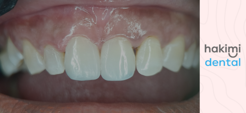
What patients sometimes describe as black on gums between the teeth are often what clinicians call black triangles: the dark, triangular spaces that appear at the base of the contacts between adjacent teeth when the interdental papilla (the gum tissue that normally fills this space) has receded.
Black triangles are not a discolouration of the gum tissue itself. They are a shadow created by the absence of gum tissue that should be present. They appear dark because the space behind them is the mouth, not because anything black is actually there.
Black triangles develop as a consequence of:
- Gum recession from periodontal disease. When the bone and connective tissue supporting the interdental papilla are lost to gum disease, the papilla follows the bone and the space emerges.
- Recession following orthodontic treatment. Teeth that have been moved through bone, particularly if moved outside the natural bone envelope, can develop recession at the interdental papilla.
- Naturally thin papilla. Some patients have naturally thin, narrow papillae that do not fully fill the interdental space, producing black triangles even in the absence of disease.
- Age-related changes. The papilla naturally recedes to some degree with age.
Managing black triangles depends on their cause. Where gum disease is responsible, professional gum treatment arrests the disease and prevents further recession, but it cannot regenerate the lost papilla in most cases. Cosmetic management of black triangles includes restorative approaches such as composite bonding to adjust the shape of the contact area between the adjacent teeth, reducing the size of the triangular space.
Amalgam tattoo: embedded metal particles
An amalgam tattoo is a localised, flat, blue-grey to black discolouration of the gum tissue caused by particles of dental amalgam (the silver-coloured metal used in older fillings) that have become embedded in the gum tissue.
This typically occurs when amalgam particles are inadvertently introduced into the tissue during a filling placement or extraction. Over time, the metal particles migrate slightly into the surrounding tissue and the sulphide compounds formed from the mercury component produce a stable, dark colouration.
Amalgam tattoos have a characteristic clinical appearance: they are flat (not raised), have irregular but generally stable borders, range in colour from grey to blue-black, and are localised to areas adjacent to existing or former amalgam restorations. They are entirely benign, do not change clinically in a way that raises concern, and require no treatment.
The clinical importance is in recognising them correctly so that they are not confused with more concerning dark lesions. Any pigmented lesion that does not clearly fit the clinical description of an amalgam tattoo, or that is adjacent to no history of amalgam use, warrants further investigation.
Acute necrotising ulcerative gingivitis: when black gums are an emergency
Black gingivitis, in its most clinically acute form, refers to what dentists call acute necrotising ulcerative gingivitis (ANUG). This is a specific, severe, rapidly progressing bacterial infection of the gum tissue that produces characteristic and unmistakeable clinical signs, one of which is a dark grey or black appearance of the necrotic (dead) gum tissue.
ANUG is quite different from chronic gum disease. It develops rapidly, over days rather than months. It is associated with:
- Intense, severe pain in the gum tissue
- Punched-out ulceration of the interdental papillae (the gum tips between the teeth), often with a grey or black necrotic slough (dead tissue) overlying them
- A very characteristic, severe bad breath that patients and those around them notice immediately
- Bleeding that occurs with the lightest contact
- A general feeling of being unwell, sometimes with fever and enlarged lymph nodes
ANUG is strongly associated with significant psychological stress, smoking, poor oral hygiene, immunosuppression (including HIV), and nutritional deficiency. It affects young adults most commonly, often students during exam periods or individuals under significant life stress.
If left untreated, ANUG progresses to acute necrotising ulcerative periodontitis (ANUP), where the necrosis extends to the bone, and then to necrotising stomatitis, where the destruction extends into the surrounding mucosa.
ANUG is a genuine dental emergency. If you or someone you know has severe gum pain, dark or black necrotic tissue between the teeth, extreme bad breath and systemic symptoms, contact our emergency dental service in Oldbury immediately. Emergency appointments at Hakimi Dental Clinic start from just £25.
The treatment is professional debridement of the necrotic tissue, irrigation with oxidising agents such as hydrogen peroxide, antibiotic therapy (typically metronidazole), appropriate analgesia, and urgent dietary and lifestyle guidance.
Medication-related gum discolouration
Several medications are associated with changes in gum colour:
- Minocycline, a tetracycline antibiotic used for acne and some infections, causes generalised dark pigmentation of multiple tissues including the gum, particularly with prolonged use. The pigment has a characteristic blue-grey quality.
- Antimalarial drugs (hydroxychloroquine, chloroquine) cause a diffuse grey-brown discolouration of the oral mucosa in some patients.
- Antifungals: Ketoconazole and some other antifungal agents have been associated with gum discolouration.
- Heavy metals: Historical exposure to bismuth, lead or silver (now very rare in clinical practice) produces characteristic grey-black lines at the gum margin, known as a bismuth, lead or silver line.
Where medication is suspected as the cause of black in the gums, the GP or prescribing clinician should be informed. In most cases the discolouration is not harmful, but it requires documentation and sometimes consideration of alternative medication.
Systemic and rare causes
A small number of systemic conditions produce gum pigmentation as part of their clinical picture.
Addison’s disease (primary adrenal insufficiency): The increased ACTH secretion that characterises untreated Addison’s disease stimulates melanocytes throughout the body. Oral hyperpigmentation, particularly on the buccal mucosa and gum tissue, is a well-recognised feature. The pigmentation may be one of the first clinical signs of the condition. Suspicion of Addison’s disease on the basis of oral findings should prompt urgent GP referral.
Peutz-Jeghers syndrome: An autosomal dominant condition producing hamartomatous polyps in the gastrointestinal tract, associated with characteristic melanotic macules (dark spots) on the lips, perioral skin and oral mucosa. These spots are typically small, discrete and multiple. This condition is important because of the associated risk of gastrointestinal malignancy.
Oral melanotic macule: A benign, localised patch of increased melanin pigmentation in the oral mucosa. Flat, well-defined, usually less than 1cm, and stable over time. Most common on the lip or palate. Clinically benign, but should be assessed and documented by a dentist.
When dark gum tissue needs urgent professional assessment
Most causes of black gums are benign. The following clinical features in any dark or discoloured area should prompt prompt professional assessment rather than watchful waiting:
- A lesion that has changed. Any dark area that has grown, changed colour, changed border definition, or developed since it was last seen needs assessment. Stable, unchanged pigmentation is reassuring; change is a warning signal.
- A raised or nodular area. Physiological pigmentation and amalgam tattoos are flat. A raised, thickened or nodular dark lesion is not typical of any of the benign causes described and needs biopsy.
- Irregular or poorly defined borders. The borders of benign pigmented lesions tend to be relatively well-defined. Irregular, poorly defined or asymmetric borders are features that raise clinical suspicion.
- Any dark lesion in a non-pigmented patient with no obvious benign explanation. A new dark lesion in a pale-skinned patient with no history of amalgam at that site and no systemic condition explaining the pigmentation needs investigation.
- Oral malignant melanoma, though rare, does occur in the oral cavity. It is most commonly found on the palate and maxillary gingiva. It presents as a dark, often irregular lesion that may be raised. The prognosis is poor when caught late and significantly better when caught early, which is why a suspicious dark lesion in the mouth should always be assessed by a qualified clinician.
The role of professional care in monitoring gum health
Whether black on gums is physiological pigmentation, smoker’s melanosis, an amalgam tattoo or something that requires further investigation, regular professional assessment through dental check-ups creates the baseline documentation that allows any changes to be detected.
A photograph or detailed clinical note of the appearance, size and location of any gum pigmentation or lesion at each visit means that any change is identifiable against an objective record. This is particularly valuable for patients with multiple or widespread gum pigmentation, where a new or changing lesion might otherwise be overlooked against an already pigmented background.
Regular dental hygienist appointments also maintain the gum health that is the context in which any dark tissue should be assessed. Healthy gum tissue surrounding a pigmented lesion behaves differently from inflamed, diseased tissue, and the two are distinguished both visually and on probing.
The bottom line
Black gums have a wide range of causes: entirely benign physiological melanin pigmentation in patients with naturally darker skin tones, smoker’s melanosis from tobacco use, amalgam tattoos from embedded metal particles, black triangles from gum recession, and at the more urgent end of the spectrum, black gingivitis in the form of ANUG, medication-related discolouration, and the rare but important possibility of a malignant lesion.
The approach is the same regardless of the cause: professional assessment at a dental check-up establishes what the discolouration actually is, documents its character and extent, and determines whether monitoring, treatment or referral is the appropriate next step.
At Hakimi Dental Clinic in Oldbury, Birmingham, dental check-ups start from £35 and emergency appointments from £25 for urgent presentations. The team is here for both.
Disclaimer
The information in this article is intended for general educational guidance only and does not constitute personalised dental or medical advice. Any new or changing area of dark tissue in the mouth should be assessed by a qualified dental professional promptly.
Hakimi Dental Clinic is a dental practice at 51a New Birmingham Road, Oldbury, B69 2JF. We offer dental check-ups from £35, emergency dental appointments from £25, dental hygiene appointments, dental implants, Invisalign, composite bonding, porcelain veneers, teeth whitening, dental crowns and smile makeovers.
FAQs
For many people, yes. Physiological melanin pigmentation produces naturally dark or black gum tissue in individuals of African, South Asian, Middle Eastern and Mediterranean heritage. This is entirely normal, benign and present since childhood. It requires no treatment. However, any new or changing area of dark tissue in anyone warrants professional assessment at a dental check-up to confirm its nature, because not all dark gum tissue is benign.
Black gingivitis typically refers to acute necrotising ulcerative gingivitis (ANUG), a severe bacterial infection producing necrotic (dead) dark tissue at the gum margins, severe pain, extreme bad breath and systemic symptoms. It is a dental emergency that needs same-day professional treatment. If you have severe gum pain with dark necrotic tissue and marked bad breath that came on over days rather than months, contact our emergency dental service immediately. Emergency appointments are available from just £25.
Yes. Smoker’s melanosis is a tobacco-related increase in gum pigmentation caused by nicotine and other tobacco compounds stimulating melanocytes in the gum tissue. It produces a diffuse brown to black discolouration, typically affecting the lower front gum tissue. The pigmentation itself is benign, but its presence indicates significant tobacco exposure, which is the primary risk factor for oral cancer. Thorough soft tissue examination at regular dental check-ups is particularly important for smokers. The pigmentation often reduces substantially within six to twelve months of stopping smoking.
Localised black spots on the gum can have several causes: amalgam tattoo (embedded metal particles from a previous filling, common near filled teeth), a melanotic macule (a benign localised dark patch), or in rare cases a nevus or other pigmented lesion requiring biopsy. A localised black spot should be assessed at a dental check-up where its location, size, border characteristics and relationship to existing restorations can be evaluated. Most are benign, but clinical assessment is the appropriate first step.
Seek same-day care through our emergency dental service if: dark gum tissue is accompanied by severe pain (possible ANUG), a dark lesion has appeared rapidly and is changing quickly, there is swelling alongside the dark tissue, or you feel systemically unwell alongside gum changes. Book a dental check-up promptly (within a week) if: a new dark area has appeared that was not there before, an existing dark area has grown or changed its character, a dark area has raised or nodular features, or a dark lesion has irregular or poorly defined borders. Stable, longstanding dark pigmentation in a patient with a clear benign explanation can be reviewed at the next routine check-up.